Visualisation of hydrogen atoms in a perdeuterated lectin-fucose complex reveals key details of protein-carbohydrate interactions
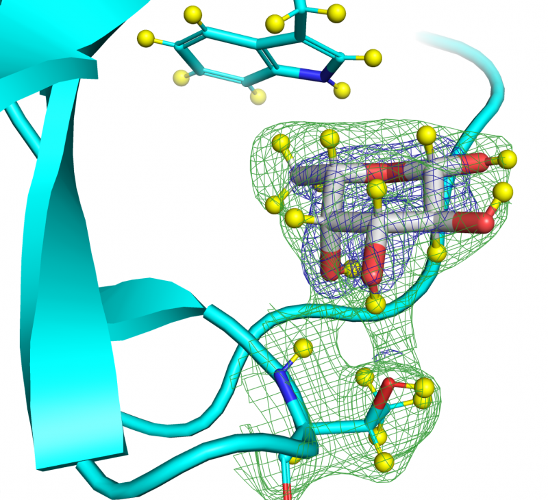
The pathogenic bacteria attach themselves to the cells which they attack thanks to the sugars present on their membrane. A crucial mechanism to study, because it would help to treat these infections without reinforcing the resistance of the bacteria. To do this, researchers from CERMAV (CNRS), the Laue-Langevin Institute (ILL) and CEITEC (Masaryk University, Czech Republic) have modified a sugar so that it responds to neutron crystallography, a technique that reveals how bacteria cling to sugars. This work, published in the journal Structure, could extend to more complex sugars to better understand different biological phenomena and propose new anti-infectious strategies. Caption: Fucose molecule in the lectin binding site. The blue grid represents the density determined by x-rays and the green grid the density determined by the neutrons around the fucose and the amino acids of the lectin. Hydrogen atoms (here isotope deuterium) are represented by yellow balls. The continuity of the green grid between the fucose and the amino acids allows a direct view of the hydrogen bonds. © L. Gajdos. / Click on the title for more information.
Replay: Serge Perez’s seminar: “Starch: An iconoclast view on amylopectin”, presented at the 7th “Special Infogest Webinar on Food Digestion”
Click on the title for more information
Small Angle Neutron Scattering Shows Nanoscale PMMA Distribution in Transparent Wood Biocomposites
« Transparent wood » can be prepared by filling the pores with resine, but how are the resine distributed. By small angle neutron scattering on samples with varyous the isotope composition of the resine we showed unambiguouslly the nanomeric ditribution of resine in the interstices of microfibrils in the cell wall. Click on the title to access more information.
Crystal and molecular structure of V-amylose complexed with ibuprofen
We have published an article in Carbohydrate Polymers about the structure of an inclusion compound prepared by crystallizing amylose in the presence of ibuprofen, a well-known anti-inflammatory drug. Using data from solid-state NMR, and electron and X-ray diffraction, we have proposed a molecular model allowing to locate the ibuprofen guest molecules in the lattice of amylose helices. Click on the title for more information.
Process-dependent nanostructures of regenerated cellulose fibres revealed by small angle neutron scattering
The nanostructure and properties of regenerated cellulose fibers depends on the spinning process. We revealed different structures by collecting small angle neutron scattering data on a series of fibers exposed to heavy water vapours. Click on the title for more information.
Daniel Marquez-Martin thesis defense on March, 16th 2021
Daniel Marquez-Martin from Cermav-UPR5301 and SyMMES- UMR5819 laboratories will defend his thesis entitled: “Versatile biosensor for deciphering glycoenzymatic activities”. Click on the title for more information.